How to Know if You Have Bradycardia

CONTENTS
- Rapid Reference 🚀
- Why bradycardia is dangerous: physiology review
- Causes
- Evaluation
- Resuscitation overview
- Medical resuscitation arm
- Atropine
- Epinephrine
- Calcium
- Other medications
- Electrical resuscitation arm
- Transcutaneous pacing
- Transvenous pacing
- Dual pacing equally a fill-in strategy
- Podcast
- Questions & word
- Pitfalls
- Supplemental media
rapid reference
(dorsum to contents)
bradycardic peri-arrest:
 pacemaker crook sail:
why bradycardia is dangerous
(back to contents)
 the effect of tachycardia on cardiac output is often overestimated
- Tachycardia has a mixed impact on cardiac output:
- Increase in heart rate tends to increase the cardiac output.
- Decreased filling time tends to decrease the stroke volume, which decreases cardiac output.
- Mild-moderate tachycardia will generally increase cardiac output. This is a normal physiologic response to stress. The issue of increased middle rate predominates here.
- Astringent tachycardia (heart rates >>150 b/k) may driblet the cardiac output because the middle doesn't have time to fill up with blood during diastole, causing a reduced stroke book.
- The deleterious effect of heart rate on cardiac output is oftentimes overestimated. For example, if a patient has atrial fibrillation with a eye charge per unit of 150 b/one thousand, it'southward unlikely that cardioversion or rate control will better cardiac output. Normally, slowing down a moderate tachycardia will cause deterioration.
the effect of bradycardia on cardiac output is often underestimated
- Bradycardia directly pulls down the cardiac output, potentially causing stupor.
- Slowing downwardly the center charge per unit may cause a minimal increase in diastolic filling, thereby increasing the stroke volume. Notwithstanding, this compensatory gene is weak and extremely limited. For example, if the heart rate decreases past a factor of two, the stroke volume cannot possibly double!
- In severe bradycardia, the cardiac output must exist low. This is simple math.
- Cardiogenic shock is divers equally inadequate cardiac output to back up organ function. Some patients tin compensate for low cardiac output without developing shock. However, with increasingly severe bradycardia, there should exist an increasing business concern for cardiogenic shock.
don't be fooled by normal-pressure bradycardia

- "the heart rate is 25 b/m merely the blood pressure is fine… I remember we tin send her to the floor"
- Some patients with bradycardia will maintain a normal blood pressure, due to an endogenous sympathetic response causing vasoconstriction. Despite a normal blood pressure, these patients notwithstanding have a low cardiac output and nevertheless may be in shock.
- Rare patients can present with astringent bradycardia and astringent hypertension (#three in figure above). Hypertension is caused by a massive sympathetic response, equally the trunk struggles to compensate for the bradycardia. This dangerous situation must exist managed thoughtfully, because the sympathetic response is really keeping the patient live. Ambitious vasodilation to treat the "hypertensive emergency" may cause hemodynamic collapse. Management should focus on correction of the bradycardia. Once the center rate normalizes, the endogenous sympathetic response should relax and everything will resolve.
progressive bradycardia is frequently a harbinger of death
- Progressively worsening bradycardia is oftentimes seen immediately preceding death ("the patient is bradying downwards").
- If the patient'south heart rate is consistently dropping in front of your eyes don't simply stand there – get some epinephrine. Fast.
- The differential diagnosis of bradycardia hither is broader than usual and may include such entities as severe hypoxemia and right ventricular failure from massive PE. Immediate evaluation should focus on the ABCs: airway, breathing, and apportionment (bedside echocardiogram).
one more reason to fear bradycardia: torsade de pointes
- Torsade de pointes is a pause-dependent arrhythmia, which is more likely to occur at slower eye rates. Furthermore, bradycardia itself may prolong the QT interval.(15974204, 21151381) It's possible that leaving patients in a severely bradycardic country may increase their risk of torsade.
common causes
(back to contents)
- Medication/intoxication:
- Beta-blocker or calcium-channel blocker.
- Central alpha-2 agonist (clonidine, dexmedetomidine, guanfacine, tizanidine).
- Cholinergic agent.
- Digoxin, antiarrhythmics (including amiodarone, dronedarone, sotalol).
- Propofol infusion syndrome.
- Alpha-blockers (e.yard. prazosin).
- Intoxication with benzodiazepines, alcohol, or opioids tin can reduce the heart rate somewhat, only this isn't normally a primary characteristic of the intoxication.
- Ketabolic:
- Hyperkalemia 📖, specially BRASH syndrome 📖.
- Hypermagnesemia.
- Hypothyroidism (myxedema coma).
- Hypothermia.
- Hypoglycemia.
- Severe hypoxia / hypercapnia / acidemia (sinus bradycardia is a common pathway of impending death from whatsoever cause).
- MI
- Neurologic catastrophe:
- Cushing's reflex due to increased ICP.
- Neurogenic shock.
- Infection:
- Lyme disease, syphilis.
- Aortic valve endocarditis with ring abscess (conduction block).
- Senile degeneration of sinus node or conduction system.
- Iatrogenic AV block:
- Following cardiac surgery (especially valvular surgery).
- Following transcutaneous aortic valve replacement (TAVR).
- Swan-Ganz catheter placement.
evaluation
(back to contents)
concrete exam
- Primary focus is the capability of perfusion.
- Overt bradycardic daze: altered mental status.
- Occult bradycardic shock: Blood pressure and mental status remain intact, but absurd extremities & poor urine output reveal inadequate perfusion.
- Cardiopulmonary exam with ultrasound:
- Book condition?
- Show of pulmonary congestion (e.g. B-lines throughout the lung fields)?
- Neuro/toxicologic exam
- Evidence of elevated intracranial pressure? (e.k. stupor, widened optic nerve sheath 🌊)
- Pinpoint pupils may advise toxic ingestion (e.g., clonidine or cholinergic agent).
 EKG: Focus on three things
- Rhythm diagnosis (e.k., sinus bradycardia vs. heart block).
- Signs of hyperkalemia (e.g., peaked T-waves).
- Signs of ischemia.
medication review
- Active medication list?
- Recent medication changes, including dose titration?
- Some medications can unexpectedly cause bradycardia (e.g. donepezil, tizanadine).(25456780) If the patient simply started a medication, expect up whether it can cause bradycardia.
- Fifty-fifty heart drops with sympatholytic properties may be enough to cause bradycardia in elderly patients.(27484658)
- Drug interactions?
- Renally cleared meds plus acute kidney injury?
labs
- Fingerstick glucose if altered mental condition.
- Chemistries including Ca & Mg.
- Troponin if MI suggested by history/EKG.
- Digoxin level, for patients taking digoxin.
- Consider checking thyroid stimulating hormone, Lyme serology.
resuscitation
(dorsum to contents)
 overview: algorithm for bradycardic peri-arrest
- Bradycardiac peri-abort may be loosely defined as severe bradycardia with marked shock and concern for immediate cardiac arrest. The algorithm below shows a maximally aggressive strategy designed to prevent further deterioration into cardiac arrest.
- At that place are two "artillery" of therapy: electrical & medical.
- It'southward hard to predict which patients volition respond best to medical or electrical therapy.
- Continue simultaneously downwardly both arms of therapy as rapidly as possible until the patient is stabilized.
- For patients with mild signs of organ malperfusion (eastward.one thousand. normal claret force per unit area but poor urine output), then a more than gradual and stepwise arroyo may exist most appropriate. For example, merely starting an epinephrine infusion will often improve heart rate and perfusion.
atropine?
(back to contents)
problems with atropine
- At low doses, atropine may cause paradoxical bradycardia.(15114081, 25634857, 16115264, 12734175)
- Atropine works by poisoning the vagus nerve, and so it is just effective for bradycardias mediated by backlog vagal tone. It will predictably neglect in cases of high-caste AV block.
- Atropine is contraindicated in patients who accept had cardiac transplantation, in whom information technology may precipitate asystole.(15114081)
- Atropine may stabilize the patient for 30-60 minutes, but then wear off. This tin can initially make the patient appear stable, but to deteriorate afterwards (one time anybody has stopped paying so much attention).
strategy when using atropine?
- If atropine is the most immediately available drug, then give it. Alternatively, if you have immediate admission to epinephrine, it may exist more effective to go straight to epinephrine.
- Atropine is traditionally the 1st-line medical therapy. However, for very unstable patients, epinephrine is more than reliably constructive and may be preferable.
- Start at one mg atropine, boosted doses can be given to a maximal dose of ~iii mg (although larger doses may be needed in patients with cholinergic poisoning).
- Overall simply ~25% of patients have a complete response to atropine, so don't delay other therapies while waiting for atropine to work.(10459592)
- Don't give atropine, sit back, and expect that it will fix everything. Give atropine while simultaneously preparing epinephrine and transcutaneous pacing – with the full expectation that the atropine will frequently neglect.
epinephrine
(back to contents)
advantages of epinephrine
- Epinephrine is available everywhere and can be obtained quickly.
- Unlike atropine, epinephrine stimulates the entire myocardium. This provides epinephrine with a broader spectrum of efficacy for various mechanisms of bradycardia.(18701603)
- Epinephrine is condom for peripheral infusion (you lot don't need to place a central line).🌊
epinephrine strategy
- Boluses for peri-arrest patient:
- For patient on verge of a cardiac arrest, bolus with doses of ~twenty-50 mcg epinephrine.
- Boluses will stabilize the patient for a few minutes, only this is only a temporary bridge to an epinephrine infusion.
- Epinephrine infusion:
- The usual dose is ii-ten mcg/min (but there is no difficult upper limit in a crashing patient).
- Dosing strategy depends on how unstable the patient is. For more than unstable patients, outset high and down-titrate as the patient responds. For patients who are adequately stable, offset depression and gradually up-titrate.
- Figure out how to achieve this at your unit:
- a) If you lot take immediate access to pre-mixed epinephrine bags, know how to use them (know their concentration and how many ml'south are needed to deliver button-dose epinephrine).
- b) If you don't have immediate access to pre-mixed epinephrine, and so, read on…
creating & using a "dirty epi drip"
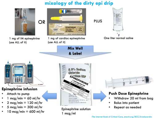 Mixing a bag of epinephrine is easy. This is conventionally termed a "dirty epi drip," but if done properly it'due south a safe and precise way to deliver epinephrine.
step #1: create the epinephrine reservoir pocketbook
- Inject ane mg of epinephrine into a liter bag of normal saline. One milligram of epinephrine can exist obtained either from an entire syringe of cardiac epinephrine (1:x,000) or an entire vial of IM epinephrine (1:1000).
- Squish around the bag to mix well.
- Label the handbag.
pace #ii: push button dose epinephrine
- For a patient in peri-abort, you volition want to deliver small boluses of epinephrine until the patient stabilizes.
- Fill up up an empty twenty cc syringe with diluted (ane mcg/ml) epinephrine from your one-liter bag.
- Bolus the patient with 20 ml of this solution, which will deliver a bolus of 20 mcg epinephrine.
- Refill your 20 ml syringe and repeat every bit needed.
- Push-dose epinephrine is a temporizing solution. As soon as the patient stabilizes, starting time an epinephrine infusion.
step #3: epinephrine infusion
- Attach your bag of epinephrine to an infusion pump and set the rate. For example:
- Infuse at 60 ml/hour to accomplish 1 mcg/min infusion
- Infuse at 240 ml/hour to accomplish four mcg/min infusion
- Infuse at 600 ml/hour to accomplish 10 mcg/min infusion
advantages of the "dirty epi" bolus & baste strategy:
- This is relatively idiot-proof. Equally long every bit you mix well and characterization the bag, it should be pretty difficult to make dosing errors:
- Regardless of what type of epinephrine you use, you lot will be fine (either ane:1,000 or 1:10,000 will piece of work).
- It'southward physically impossible to bolus a lethal dose of epinephrine later it's been diluted to i mcg/ml (yous would need a >100 ml syringe, which doesn't be).
- Even if you run the epinephrine purse in wide open up, y'all would merely be delivering about ~30 mcg/min of epinephrine – and then over again, it's basically impossible to evangelize a lethally high epinephrine dose.
- Constructing a reservoir purse of epinephrine encourages a rapid transition from push-dose epinephrine to an epinephrine infusion (which is ultimately a safer and more than controlled strategy).
- This is a viable approach during epinephrine shortages:
- Easily performed with 1:1000 epinephrine, if your shop runs out of 1:ten,000 epinephrine.
- Ane vial of epinephrine can be used for both pushes & drip, thereby conserving medication.
calcium
(back to contents)
Along with epinephrine, calcium is a drug which is often under-utilized in bradycardia. IV calcium is potentially effective for various etiologies listed below. Calcium is pretty safe (unless information technology extravasates), and so when other therapies neglect it makes sense to try to some calcium.
calcium-responsive bradycardias:
- Hyperkalemia.
- Hypocalcemia.
- Hypermagnesemia.
- Calcium-channel blocker.
- Beta-blocker (maybe).
dosing
- Bradycardia of unknown etiology: Try one round of calcium (ane gram calcium chloride or 3 grams calcium gluconate).
- Known or suspected hyperkalemia: Start with 1 gram of calcium chloride or 3 grams of calcium gluconate. If ineffective and patient is dangerously unstable, consider additional calcium. The maximal dose of calcium is unknown in this state of affairs. Bedside chemical science monitoring with an iSTAT might be helpful here, shooting for moderate hypercalcemia (e.1000., an ionized calcium level of 2-3 mM).
other medications
(dorsum to contents)
dobutamine
- Dobutamine is more often than not a beta-agonist, with very weak alpha-adrenergic activity. Different epinephrine, dobutamine tends to cause systemic vasodilation:
- Dobutamine might exist perfect for a patient with bradycardia and normal/elevated blood pressure, where you're trying to increase cardiac output (without increasing the blood pressure).
- Dobutamine isn't a good choice for the crashing, hypotensive patient. If the dobutamine fails to accelerate the heart rate then information technology could human activity solely every bit a vasodilator, and thereby cause worsening hypotension.
- Dobutamine might non be quite every bit safe for peripheral infusion every bit epinephrine. If you're giving dobutamine for prolonged peripheral infusion, monitor the site advisedly and avert any IVs in the manus or wrist.
isoproterenol
- This is an excellent drug for bradycardia if you can become ahold of it.
- Isoproterenol is a pure beta-agonist, which is safe for peripheral infusion. Isoproterenol does seem to exist a flake more powerful than epinephrine (there are some patients who don't respond to epinephrine all the same will answer to isoproterenol).
- The main drawbacks to isoproterenol are logistic. Isoproterenol is insanely expensive in the The states (an infusion may price several grand dollars). Many hospitals don't have it. Even if your hospital does have information technology, information technology will usually take time getting it from pharmacy.🌊
dopamine
- Dopamine has a long runway record of use in symptomatic bradycardia. The chief advantage of dopamine is that it'southward stable at room temperature, so it may be more widely available in pre-mixed bags (e.chiliad., in ambulances).
- Disadvantages of dopamine compared to epinephrine:
- 1) Dopamine can cause skin necrosis with prolonged infusion.
- 2) At high doses, dopamine may act predominantly every bit a vasoconstrictor. This tin be undesirable if you're by and large looking for chronotropy.
- If dopamine is the most readily available agent, and so apply it. When yous accept time, consider switching over to an epinephrine infusion.
aminophylline 💊
- May occasionally be useful in certain situations, including:(30586772, 31311698)
- Patients status post cardiac transplant.
- Second- or 3rd-degree AV block associated with acute junior MI.
- Spinal cord injury.
- Carotid sinus syncope. 📄 (34316890)
- Ticagrelor induced bradycardia.(27920235)
- The usual loading dose is 6 mg/kg platonic body weight, infused over 20-30 minutes. If effective, this may be followed by a maintenance infusion of aminophylline (e.g., 0.iii-0.five mg/kg/hour) which may somewhen be transitioned to chronic oral maintenance theophylline. The optimal dosing of aminophylline/theophylline for bradycardia is unknown. Some case reports advise that relatively low doses may be effective, but it may too exist reasonable to target a theophylline level of ~10-20 ug/ml (like to its use in obstructive airway disease). 📄 (18154483, 34316890)
avant-garde toxicologic therapies
- Local Anesthesia Systemic Toxicity (LAST) 📖
- Suspect in any bradycardic patient on lidocaine infusion or recently treated with nerve cake.
- Front-line therapy is intralipid.
- Beta-blocker and/or calcium-aqueduct blocker toxicity 📖
- Advanced toxicologic treatments are primarily useful for patients who nowadays with massive overdose. Nonetheless, these therapies can also be considered for patients with bradycardia due to therapeutic misadventures.
- Handling may involve high-dose insulin, glucagon, or intralipid.
transcutaneous pacing
(dorsum to contents)
Transcutaneous pacing is often the fastest strategy to increase the heart rate. Even if it doesn't capture, the discomfort may be plenty to trigger a sympathetic response that keeps the patient alive. Either mode, this is a temporary measure until more definitive stabilization is possible (e.thousand., transvenous pacing).
 pad configuration
- Air is a poor usher of electricity, so placing pads that overlie the lungs is a poor strategy.
- Anterior-posterior pad placement may be preferred (epitome above)(26849986)
- Anterior pad is on the left side of the lower part of the sternum, covering the "left parasternal window" of the heart. Based on experience with echocardiography, this is the almost reliable site of contact between the eye and the soft tissue of the chest.
current
- If patient is crashing, start at maximal current and piece of work your way down after the patient has stabilized.
- If patient is doing OK, so start depression and titrate up.
- If the patient is doing OK, then you probably wouldn't really want to do transcutaneous pacing at all. Still, it may be useful to determine if the patient responds to transcutaneous pacing. Proving that transcutaneous pacing will capture the heart may help y'all decide whether placing a transvenous pacemaker is necessary in a borderline patient.
- Continue pacing at 10-20 mA above the minimum energy required for capture.
- Unremarkably ~40-80 mA required to achieve capture (possibly more than in obesity or obstructive lung disease).(24044868)
beware of pseudo-pacing

- Pseudo-pacing is when the pacemaker isn't capturing the myocardium, only the monitor displays a middle rate equal to the transcutaneous pacemaker. This provides a false sense of security, considering the monitor looks bang-up.
- Ever confirm that the pacemaker is capturing via i of the following methods:
- Pulse oximetry waveform shows a pulse matching the pacemaker (epitome in a higher place).
- Bedside echocardiogram confirms myocardial contraction with pacing.
- Pulse, preferably far away from the breast (e.yard., femoral pulse or dorsalis pedis – to avert existence fooled by twitching of the breast musculature).
analgesia/sedation?
- This tin be limited by patient'due south instability. Low-dose fentanyl and/or ketamine might be reasonable.
- Deep sedation & intubation to allow for tolerance of transcutaneous pacing is a popular approach, but probably not the best. The instability induced by sedation and intubation may outweigh benefits from transcutaneous pacing. Also, if the patient becomes hyperinflated on the ventilator, this could theoretically lead to loss of capture by the transcutaneous pacer.
transvenous pacing
(back to contents)
Transvenous pacing is the nigh invasive strategy, but likewise the most effective (with success rates >95%).(17212976) Indications are roughly as follows:
- Unstable bradycardia which doesn't respond to other interventions (e.g., epinephrine).
- High-degree AV blocks that leave the patient at ongoing hazard of deterioration (east.g., Mobitz II, third-degree heart cake with wide-complex escape rhythm).
take a kit, know your kit, love your kit
- The unit should accept everything needed for a transvenous pacer in ane specific location (eastward.g., a large box or drawer in a resuscitation cart). This will include the transvenous pacemaker itself, the venous sheath, the pacing generator, wires, and adapter pins.
- Use the appropriate size venous sheath for the pacemaker:
- If yous ask for a random venous sheath, you're likely to be given an 8.5 French sheath which is designed to accommodate a Swan-Ganz catheter. This sheathcannot be used for placement of a pacemaker wire – it will be too big (leading to leakage of blood out of the sheath or air embolization into the sheath). The pacer sheath will be smaller.
- The crux of this process is familiarity with the pacer kit stocked in your unit of measurement. Ideally the unit should have a not-sterile kit available for training purposes. In an emergency, the muscle memory for how all the parts get assembled volition be invaluable.
- Also know how to piece of work the pacing generator. Newer digital pacing generators are designed for electrophysiologists, and then they tin be confusing. Brand sure you lot're familiar with your hospital's device.
 insertion site
- In full general, the sites which allow for most facile floating of the pacemaker are:
- 1st choice: Right internal jugular (directly shot into the RV).
- 2nd choice: Left subclavian (smoothen arc through the larger vessels into the heart).
appropriate electric current while floating the wire
- Depending on how unstable the patient is, there are roughly two strategies for floating a temporary pacemaker:
- Dear Badger Style : Every bit you lot are floating the wire, increment the electric current to 20 mA. This will capture the heart as apace every bit possible, which is preferable if the patient is actively dying. The problem is that capture may occur while the wire is in the atrium, so this arroyo doesn't ever result in ideal placement of the temporary pacemaker. The goal here is to stabilize the patient every bit before long as possible, you tin fiddle with the pacemaker later.
- Usual technique: For a patient who isn't actively dying, float the pacemaker with a lower amplitude (due east.m., v mA). This normally won't capture the myocardium until you're close to the right ventricular myocardium. This strategy is better for optimizing the platonic placement of the pacemaker. After yous gain capture, advance the pacer a couple of millimeters farther and deflate the balloon – this will often position it optimally, lying against the correct ventricle.
ultrasound guidance
- This isn't necessary, just tin be helpful. Ultrasound requires a second operator to reach nether the sterile drape and position the ultrasound.
- A four-chamber view (eastward.g., subcostal 4-chamber) is generally all-time, as this can permit visualization of the wire entering the right atrium and ventricle.
- Potential value:
- (1) If you advance the pacer wire over ~30 cm anddon't see it in the right atrium, then the wire has probably passed straight into the the junior vena cava. Deflate the balloon, pull back to ~xv cm, and try floating once again.
- (2) Ultrasound allows fine-tuning of the placement process. For example, in one case you're through the tricuspid valve you can irksome down – y'all just have a few more centimeters left to become.
- (three) If y'all visualize the wire in the right ventricle but you lot're non getting capture then there might exist a problem with the pacer box. Make sure all the wires are continued correctly and the settings are correct.
complications
- Most complication chronicle to placement of the pacemaker sheath in the vein (eastward.yard., pneumothorax or bleeding).
- There is a very minor, notwithstanding finite take chances of hemopericardium (<0.vi%) which tin can pb to tamponade 📖.(30543806) If a patient deteriorates following placement of a temporary transvenous pacemaker, ultrasonography should exist performed to exclude hemopericardium.
dual pacing every bit a fill-in strategy
(back to contents)
Some patients will exist encountered who are completely pacemaker-dependent (they accept no intrinsic charge per unit at all). This is pretty scary, because if electric pacemaking fails for fifty-fifty a minute the patient will have a cardiac arrest. Temporary transvenous pacemakers do occasionally become dislodged, and then this can be a real problem. To prevent the patient from dying if their temporary transvenous pacemaker falls out of place, dual pacing can exist used:
- The patient is simultaneously attached to both a transvenous pacemaker and a transcutaneous pacemaker.
- The transvenous pacemaker is adjusted the way it normally would exist:
- Set the rate to something reasonable (e.one thousand 60-80 b/m, or perchance college if the patient is otherwise shocky).
- This should exist the pacemaker which is driving the patient's middle charge per unit.
- The transcutaneous pacemaker is attached and turned on, with the following settings:
- Set the rate 20 b/m beneath the transvenous pacemaker (e.g. 40 b/one thousand).
- Set the current to a high enough level to capture the myocardium.
- Optimize pad placement.
- This pacemaker should ideally practise nothing.
The transcutaneous pacemaker is used here purely as a backup device. If the transvenous pacer malfunctions, the transcutaneous pacemaker will pick up without losing a beat. Of course, this volition be painful for the patient (considering all of a sudden they volition exist getting shocked). Still, transcutaneous pacing is preferable to sudden cardiac arrest.
podcast
(dorsum to contents)

Follow us on iTunes
questions & discussion
(back to contents)
To keep this page small and fast, questions & discussion about this post tin can exist found on another page here .

- Don't assume that because the claret force per unit area is normal, the patient is fairly perfusing and doing fine. Some patients vasoconstrict and maintain normal blood pressure, despite organ malperfusion.
- For an unstable patient, don't get fixated on whatever specific intervention. Proceed working through a serial of electric and mechanical therapies until something works (figure below).
- Don't exist agape to use push button-dose epinephrine and peripheral epinephrine infusions for an unstable patient.
- Don't forget to go a skilful medication history, focusing on recent medication changes and drugs which can accumulate in renal dysfunction (e.m. digoxin, atenolol).
- Don't be fooled by transcutaneous pacemaker pseudocapture. The fact that the breast is twitching and the monitor shows a normal center rate ways nothing – it'southward still possible that the myocardium isn't being captured.
- Recollect that bradycardia can be caused past myocardial infarction and various intoxications – so fixing the heart rate may not be enough to fix the patient.
- Attempt to imagine every piece of your transvenous pacemaker kit and how they it is assembled. If you lot can't do this, you need practice with the kit. The nigh common procedural hang-upwardly is beingness unfamiliar with the kit and pacemaker generator.
Guide to emoji hyperlinks
Going further
- Bradycardia
- Atropine vs. Epinephrine for bradycardic periarrest (PulmCrit)
- Bradycardia (Chris Nickson, LITFL)
- Managing unstable bradycardia (First 10 EM, Justin Morgenstern)
- Symptomatic Bradycardia (Erica Simon, emDocs)
- An arroyo to bradycardia in the emergency department (Patrick Ng, emDocs)
- BRASH syndrome & failure of the ACLS bradycardia algorithm (PulmCrit)
- Dirty Epi Drip & Push Dose Epi
- Push dose pressors & Push-dose pressor update (EMCrit)
- Muddied Epi Drip (Zlatan Coralic, ALIEM)
- Transcutaneous pacing
- Transcutaneous pacing success & false capture (EMS 12-lead)
- Transcutaneous pacing (Chris Nickson, LITFL)
- Transvenous pacemaker insertion
- Transvenous pacing: Dr. Smith's ECG blog
- Temporary pacemaker troubleshooting & Transvenous pacing (LITFL)
- Transvenous Pacemaker Placement (Taming the SRU)
- Transvenous Pacing (John Sarwark, EM Curious)
- The Beloved Annoy
- This is essential learning.
supplemental media
(back to contents)
#POCUS dx? pic.twitter.com/r1MFw7ekQ4
— Philippe Rola (@ThinkingCC) April 6, 2018
References
- 10459592 Brady WJ, Swart K, DeBehnke DJ, Ma OJ, Aufderheide TP. The efficacy of atropine in the treatment of hemodynamically unstable bradycardia and atrioventricular cake: prehospital and emergency department considerations. Resuscitation. 1999 Jun;41(one):47-55. doi: x.1016/s0300-9572(99)00032-5 [PubMed]
- 12734175 Maruyama Grand, Mochizuki North, Hara K. High-degree atrioventricular block afterward the administration of atropine for sinus arrest during anesthesia. Can J Anaesth. 2003 May;50(5):528-9. doi: 10.1007/BF03021079 [PubMed]
- 15114081 Bernheim A, Fatio R, Kiowski W, Weilenmann D, Rickli H, Brunner-La Rocca HP. Atropine often results in complete atrioventricular block or sinus abort afterward cardiac transplantation: an unpredictable and dose-independent miracle. Transplantation. 2004 Apr 27;77(8):1181-5. doi: 10.1097/01.tp.0000122416.70287.d9 [PubMed]
- 15974204 Ashworth SW, Levsky ME, Marley CT, Kang CS. Bradycardia-associated torsade de pointes and the long-QT syndromes: a case report and review of the literature. Mil Med. 2005 May;170(5):381-half dozen. doi: 10.7205/milmed.170.5.381 [PubMed]
- 16115264 Chin KJ, Seow SC. Atrioventricular conduction block induced by low-dose atropine. Amazement. 2005 Sep;60(nine):935-6. doi: 10.1111/j.1365-2044.2005.04346.x [PubMed]
- 17212976 Sodeck GH, Domanovits H, Meron K, Rauscha F, Losert H, Thalmann Yard, Vlcek M, Laggner AN. Compromising bradycardia: management in the emergency department. Resuscitation. 2007 April;73(1):96-102. doi: x.1016/j.resuscitation.2006.08.006 [PubMed]
- 18154483 Whitman CB, Schroeder WS, Ploch PJ, Raghavendran Chiliad. Efficacy of aminophylline for handling of recurrent symptomatic bradycardia afterwards spinal cord injury. Pharmacotherapy. 2008 Jan;28(i):131-v. doi: 10.1592/phco.28.1.131 [PubMed]
- 18701603 Vavetsi S, Nikolaou N, Tsarouhas G, Lymperopoulos G, Kouzanidis I, Kafantaris I, Antonakopoulos A, Tsitsimpikou C, Kandylas J. Consecutive administration of atropine and isoproterenol for the evaluation of asymptomatic sinus bradycardia. Europace. 2008 Oct;ten(10):1176-81. doi: 10.1093/europace/eun211 [PubMed]
- 21151381 Namboodiri Due north. Bradycardia-induced Torsade de Pointes – An arrhythmia Less Understood. Indian Pacing Electrophysiol J. 2010 Oct 31;ten(ten):435-8 [PubMed]
- 24044868 Deal N. Evaluation and direction of bradydysrhythmias in the emergency department. Emerg Med Pract. 2013 Sep;15(9) [PubMed]
- 25456780 Cortes J, Hall B, Redden D. Profound symptomatic bradycardia requiring transvenous pacing after a unmarried dose of tizanidine. J Emerg Med. 2015 Apr;48(4):458-threescore. doi: 10.1016/j.jemermed.2014.10.005 [PubMed]
- 25634857 Carron M, Veronese Due south. Atropine sulfate for treatment of bradycardia in a patient with morbid obesity: what may happen when you least expect information technology. BMJ Case Rep. 2015 Jan 29;2015:bcr2014207596. doi: x.1136/bcr-2014-207596 [PubMed]
- 26849986 Seifert PC, Yang Z, Reines HD. Crisis Management of Unstable Bradycardia in the OR. AORN J. 2016 Feb;103(ii):215-23. doi: 10.1016/j.aorn.2015.12.012 [PubMed]
- 27484658 Wung SF. Bradyarrhythmias: Clinical Presentation, Diagnosis, and Management. Crit Intendance Nurs Clin North Am. 2016 Sep;28(3):297-308. doi: ten.1016/j.cnc.2016.04.003 [PubMed]
- 30543806 Metkus TS, Schulman SP, Marine JE, Eid SM. Complications and Outcomes of Temporary Transvenous Pacing: An Analysis of > 360,000 Patients From the National Inpatient Sample. Chest. 2019 Apr;155(4):749-757. doi: 10.1016/j.chest.2018.11.026 [PubMed]
- 31311698 Sidhu S, Marine JE. Evaluating and managing bradycardia. Trends Cardiovasc Med. 2020 Jul;thirty(5):265-272. doi: ten.1016/j.tcm.2019.07.001 [PubMed]
Source: https://emcrit.org/ibcc/bradycardia/
Enregistrer un commentaire for "How to Know if You Have Bradycardia"